Zoologger: The Largest Arthropod to Prowl the Land: Samantha Hill

Among the beautiful beaches and gigantic waves in Palmyra atoll, just south of Hawaii, a big creature waits his chance to emerge as the sun goes down.

Jawless Vertebrae Had the Worlds Sharpest Teeth: Meghan Skura

The Conodont is recorded to have the sharpest teeth of any animal that ever lived, hoever its teeth are barely thicker than a human hair.

Ancient Creature was a Miniture Monster: Christina Vilkhovoy

A 500 million year old monster looking predator was discovered in the deposits of Canada.

Some Echinoderms Will Never Grow Up: Erica DeMello

In life, people think of being “immature” or not growing up as a bad thing. But in this case, for species like echinoderms, it can be advantageous.

500-Million-Year-Old Sea Creature Unearthed: Desiree Villeneuve

Scientists have discovered a 520-million-year-old preserved fossil of an arthropod known as a fuxhianhuiid.

Paradox Worm' Xenoturbella Bocki Lacks Brain and Sex Organs, But Could Be Mankind's 'Progenito:Dana Hebert

There is a high possibility that a paradox worm can be more related to humans that many other more advanced organisms.

Tuesday, May 7, 2013

A Look at the World Explains 90 Percent of Changes in Vegetation

photo courtesy of nasa.gov

Human kind runs this world, building, expanding and surfacing as much of mother earth as they can. Some say this is our way of flourishing in our environment and taking advantage of things earth has to offer but we must take a step back and look at the damage being done. “In the last thirty years, vegetation has changed significantly the world over (Schaepman).”

Climate is the main thing that control vegetation, being the start of the growth process and human kind can control the climate. With our excessive amounts of abuse of the environment we are not allowing earth to naturally control and regulate its self. “Within the last thirty years, for instance, vegetation activity has increased in the northern hemisphere but declined in the southern hemisphere (Schaepman).” Over all there have been drastic changes in the ecosystem due to humans activity, from driving cars to desecrating forests and it is only worsening. If nothing is done about these changes it is not known exactly what will happen but predictions of worsening living conditions have been made.

“The majority of the changes – more than 30 percent overall – were caused by human activity (Schaeoman).” This article talks about the largeness of our effect of the environment, and gives insight to just how bad it is and could be. With this information known and public it’s time to start changing out ways.

Future of organs? Synthetic tissue built with 3-D printer

photo courtesy of trbimg.com
On April 4th, 2013, Amina Khan wrote an article in the Los Angeles Times describing some new developments in the scientific and medical world. The article, entitled, “Future of organs? Synthetic tissue built with 3-D printer,” describes how scientists have developed a 3-D printer that creates materials that very closely resemble human tissues. The substances used to make this material are actually quite simple. They are networks of water droplets coated in lipids, and will someday be used to replace damaged tissue in living organs. 

The droplets of water consist of lipid bilayers, similar to cell membranes, that allow things to go in and out. Before creating the 3-D printer, this process of creating these water droplets was a tedious and laborious process. Now, since the production of the 3-D printer, it uses a micropipette to squeeze the droplets out, which sped up the process incredibly.

Scientists were surprised by these results. They did not expect that they would be able to use these droplets as tissues. The did not expect that once they could print the droplets that they would so closely resemble human tissues, and also did not expect that they would be able to make them in such an efficient way. Along with replacing human tissue, the synthetic tissue will be able to act as nerve pathways, triggered into contracting like a muscle, and even able to send electrical signals, like a nerve. These tissues will be used to graft onto organs to replace damaged parts, and to grow more cells.

Blog Post Author Taylor Berube from Section 124-28

Works Cited
4, Amina Khan April. "Future of Organs? Synthetic Tissue Built with 3-D Printer." Los Angeles
Times. Los Angeles Times, 04 Apr. 2013. Web. 19 Apr. 2013.

Tissue engineering: Growing new organs, and more

photo courtesy of mit.edu
Tissue engineering research has progressed from using tissue for replacement organs and transplants to focusing on creating tissue that can be used to study diseases and develop new drugs for treatments. Scientists at MIT are especially focused on this research. Sangeeta Bhatia, a professor at MIT, has developed a liver tissue, which has allowed her to study malaria and hepatitis c. She has used thin pieces of this liver tissue to implant in mice, which has allowed her to study new possible treatments.

In addition, scientists at MIT are in the process of developing a “human-on-a-chip” system. This would be a system of interconnected tissue that could be modified based on what diseases are being researched. This “human-on-a-chip” system would allow scientists to study multiple diseases at once and test the effects of different drugs on tissues.

Other tissue engineering research includes, research to develop regenerative therapies that would aid in healing wounds and tissue injuries. A professor at MIT, has developed implantable scaffolds that have endothelial cells within them. Endothelial cells are cells that secrete proteins that respond to injury. These cells could be used to help repair damage caused by surgeries, cancer, smoke, and cardiovascular disease. These implantable scaffolds are currently used in clinical trials to help heal blood vessel injuries that are caused by the needles used in dialysis. If these trials are successful, the implantable scaffolds could potentially double the amount of time a person with kidney failure is allowed to be on dialysis.

Lastly, professors at MIT are attempting to develop cardiac tissue that would help heal patients who have lost their voices. The cardiac tissue would include electronic sensors and a synthetic polymer. It would heal people who have lost their voices by restoring the function of their vocal cord. The challenge that arises in developing organs is that it is necessary for the blood vessels in the tissue, when implanted, to connect to the patient’s blood supply. Scientists at MIT are trying to combat this problem by inducing blood vessels to form by trying to grow cells on a nano-patterned surface.

Blog Post Author: Mary Catherine Maher 124-28

Article Citation:
Trafton, Anne. "Tissue Engineering: Growing New Organs, and More." MIT's News Office. Massachusetts Institute of Technology, 14 Dec. 2012. Web. 17 Apr. 2013

Sunday, April 14, 2013

Paradox Worm' Xenoturbella Bocki Lacks Brain & Sex Organs, But Could Be Mankind's 'Progenito

Photo courtesy of sciencedaily.com
The article “’Paradox Worm' Xenoturbella Bocki Lacks Brain & Sex Organs, But Could Be Mankind's 'Progenitor'” by Andres Jauregui of Huffington Post, describes the high possibility that a paradox worm can be more related to humans that many other more advanced organisms. This organism might belong to a branch of the animal family tree called deuterostomes. “Deuterostome development in animals, is a developmental mode distinguished by the development of the anus from the blastopore; often also characterized by radial cleavage and by the body cavity forming out pockets of mesodermal tissue” (Campbell 661). For example, the study’s co-author Matthias Obststates states, “So maybe we're more closely related to the Xenoturbella bocki worm, which doesn't have a brain, than we are to lobsters and flies” (Jauregui 1). Scientists have good reason to believe that the human race is more closely related to something so simple and far from complex as this worm that lacks a brain and sex organs! They have found that, “…the worm's early embryonic development is similar to that of humans, which could help answer questions about how human organs are formed” (Jauregui 1). This discovery could potentially help scientists regenerate organs, tissues and contribute to stem cell research. This new found information can benefit the future of medicine and longevity immensely.

Blog Post Author: Dana Hebert, Section 124-28

Article Citation

Jauregui, Andres. "'Paradox Worm' Xenoturbella Bocki Lacks Brain & Sex Organs, But Could Be Mankind's 'Progenitor'" The Huffington Post. TheHuffingtonPost.com, 28 Mar. 2013. Web. 02 Apr. 2013.

Reece, Jane B., Neil A. Campbell, Michael L. Cain, Steven A. Wasserman, Peter V. Minorsky, and Robert B. Jackson. Campbell Biology. 9th ed. Boston: Benjamin Cummings / Pearson Education, 2011. Print.

Jawless Vertebrae Had the World's Sharpest Teeth


Photo courtesy of rhyniechert.com
 This article was about a vertebra named a Conodont. It is recorded to have the sharpest teeth of any animal that ever lived. Its teeth are barely thicker than a human hair; however their strength can compete with the strength of our human jaw. This vertebra evolved 500 million years ago and went extinct about 200 million years ago. This vertebra roamed the earth for longer than any other vertebra so far. They do not have a jaw; however they were the first vertebra to evolve teeth. Conodont’s teeth are a little bit different than mammalian teeth. Instead of going up and down, the Conodont’s teeth go left to right to work. There was a problem with having such thin sharp teeth, because they’re so thin and fragile they are more likely to break. To overcome this, the Conodont was able to re-sharpen and repair their teeth throughout their lives. The discovery of these vertebra gave rise to insight of the dental evolution of vertebra. The fossilized teeth are found abundantly in the sedimentary rock in marine environments. By looking at these fossils it can be determined how vertebra evolved and what were the reason for them to evolve, which was most likely food. The finding of Conodonts opened doors for scientific investigation.

Blog Post Author: Meghan Skura, Section 124-21

Article Citation:
Bell, Alexandra. "Jawless Vertebrate had world's sharpest teeth." Nature. N.p., 14 Mar 2012. Web. 14 Apr 2013. <http://www.nature.com/news/jawless-vertebrate-had-world-s-sharpest-teeth-1.10211>.

Some Echinoderms Will Never Grow Up

photo courtesy of deepseanews.com
In life, people think of being “immature” or not growing up as a bad thing. But in this case, for species like echinoderms, it can be advantageous. Xyloplax is an example of an Echinoderm that has adapted to its preferred environment. The article goes on to name list the five living examples of Echinoderms and some examples of each. When Xyloplax was first discovered, it apparently did not fit into any of the five classes because it did not have all the characteristics of one in particular. For example, making it unique, the Xyloplax does not have arms; however it was then found out that it was most closely relate to the family Pterasteridae. It has also been hypothesized that Xyloplax is “an actual Asteroid with arrested development so that sexually mature individuals occur in an otherwise juvenile body” (Dr. M.). In other words, Xyloplax’s characteristics allow it to live in pieces of wood on the seafloor. Because it is so small, it can fit into tiny spaces in the wood and eat bacteria. The moral of the article is that next time you try and tell someone to grow up, think first about how they may be perfect the way they are!

Blog Post Author: Erica DeMello, Section 124-28

Article Citation
Janies, D., J. Voight, and M. Daly. "Some Echinoderms Will Never Grow Up." Deep Sea
     News. N.p., n.d. Web. 9 Apr. 2013.

Ancient Creature was a miniature monster


Photo courtesy of http://palaeos.com
A 500 million year old monster looking predator was discovered in the deposits of Canada. The first clues of the monster fossil were described 100 years ago and scientists thought it to be a crustacean animal, however, now its clear that Hurdia is a large predatory animal, half a meter in length with a segmented body and a head with spiny claws and a circular jaw with teeth. It is extremely rare to find a complete fossil because the soft tissues decay quickly.

More and more clues of Hurdia are accumulated over the years but the final clue was discovered when a sample turned up in storage cabinets at the Smithsonian National Museum of Natural History in Washington, D.C. This specimen has been intact until researches in the 1970’s classified it as an arthropod and then as an unusual specimen of the famous monster predator Anomalocaris. Hurdia is related to this other beast, but one thing that was different from both is that a large three-part shell projects out from the front of the animal’s head. Researchers were astonished by this structure because it was unlike anything they had seen in arthropods. The shell structure in Hurdia did not seem to cover or protect the body as most do and it was empty.

No one knows what these predators might have ate because there is no direct evidence but they think that it ate whatever came around. If it was looked at in terms of the Cambrian marine world, it may have ate marine worms, trilobites, other arthropods, molluscs, or other predators. Hurida was covered in gills which my hint it was necessary to provide oxygen to a large swimming animal. Hurdia and Anomalocaris are early lineages that directed to arthropods because they had compound eyes and limbs with filaments used for breathing.

Blog Post Author: Christina Vilkhovoy, Section 124-22


Article Citation
LiveScience Staff. “Ancient Creature was a Minature Monster.” Live Science. 19 March 2009. Web. < http://www.livescience.com/3416-ancient-creature-miniature-monster.html>.

500-million-year-old sea creature unearthed

Photo Courtesy of nbcnews.com

Scientists have discovered a 520-million-year-old preserved fossil of an arthropod known as a fuxhianhuiid. This is one of the earliest animal fossils discovered, however it is not the first fuxhianhuiid fossil discovered but it is the best example of this arthropod because it shows the internal organs. This arthropod displays primitive limbs under its head and a nervous system that goes beyond the head, which is the earliest example of a system like that.

These creatures lived during the beginning of the Cambrian explosion, 50 million years before animals had even moved from living in the sea to living on land. Scientists believe that these creatures were ones that would be found along the sea bed and believe they might have been able to swim for short distances. These creatures most likely evolved from worms with legs.

This has been a very important discovery because it may be able to help put together evolutionary history of animals. This has also been helpful because arthropods are often classified based on their head appendages and this fossil shows the earliest examples of that. "These fossils are our best window to see the most primitive state of animals as we know them – including us," Ortega-Hernández said in a statement. "Before that there is no clear indication in the fossil record of whether something was an animal or a plant – but we are still filling in the details, of which this is an important one" (Ghose).

Blog Post Author: Desiree Villeneuve, Section 124-21

Article Citation
Ghose, Tia. "500-million-year-old Sea Creature Unearthed." NBC News. NBC News, 27 Feb.
2013. Web. 05 Apr. 2013. <http://science.nbcnews.com/_news/2013/02/27/17119273-500-million-year-old-sea-creature-unearthed?lite>

Zoologger: The Largest Arthropod to prowl the land


Among the beautiful beaches and gigantic waves in Palmyra atoll, just south of Hawaii, a big creature waits his chance to emerge as the sun goes down. The creature is known as the Coconut Crab, species Birgus latro, and it is Earth’s largest land arthropod. The beast’s measurements are shocking with length up to 40 centimeters and a leg span of 90 centimeters. Although most of its other large competitors live in the ocean, the Coconut Crab has adapted a brilliant way of living.

The crabs are born at sea, usually on a new moon, when their mothers release their fertilized eggs. The newborns live in the sea for about a month and then migrate onto land. In order to be able to survive on land, Coconut crabs developed organs called branchiostegal lungs. They are basically gills turned inside out. These specialized organs were developed because their abdomens were allowed to be exposed to air, unlike other hermit crabs.

Their name implies that they feed on coconuts and many other nuts but using their claws to crack open the shell. They often carry a coconut to the top of a tree and drop it in order to crack it open. Occasionally, they steal food from campers, giving them the nickname robber crabs. Mouths are absent in these crabs, instead they have a gastric mill that grinds the nuts so they can be swallowed.

The large crabs have been hunted to their point where they are non-existent on some islands. Trying to make is safe for them to produce is hard due to the fact that the land and sea would both need to be safe. IN the lab, they have had success in all steps of breeding. Much work has to be done before these trials will be done in the wild. They are sure to help the big crab back into the world.

Blog Post Author: Samantha Hill, Section 124-23

Article Citation:
Marshall, Micheal. “Zoologger: The largest arthropod to prowl the land.” Newscientist. 03 March 2010.<http://www.newscientist.com/article/dn18600-zoologger-the-largest-arthropod-to-prowl-the-land.html>

Sunday, March 31, 2013

Reproduction and Lifespan are intertwined

An organ-shaping gene has been found in the microscopic worm Caenorhabditis elegans, or C. elegans for short. The gene and the protein it makes are responsible for shaping the gonad, a complex reproductive organ. Similar genes and proteins are probably at work in other animals, including humans. The new finding by University of Wisconsin-Madison biochemist Judith Kimble and graduate student Robert Blelloch advances the prospect of one day growing complete organs for transplant.
Photo courtesy of Judith Kimble; news.wisc.edu

The gonad is well known for being helpful with reproduction but at the same time it has cut animals life spans down. Planck just recently discovered though that there is a molecular switch that can extend an animal’s life span using components of a developmental clock (Reproduction and Life Span Are Intertwined). The commonly used organism for research is called Caenorhabditis elegans. The Caenorhabditis elegans develops throughout four stages based on the developmental clock. “Yidong Shen and others worked with the Caenorhabditis elegans and used lasers to remove the germ cells, they found that remaining cells trigger the production of a hormone called Dafachronic acid (Reproduction and Life Span Are Intertwined).”

Dafachronic acid activates microRNAs, which work as little molecular switches causing changes in gene expression that promote longer life span. Losing the germ cells causes the developmental clocks to be put in motion leading to a longer life. I found this article intriguing because I never realized that research like this was going on, and that we have to ability to do such a research. It makes me want to continue looking up articles based on the theme of each lab to keep educating myself.

Blog Post Author: Paige Brandsdorfer Section 124-26

Works Cited
"Reproduction And Life Span Are Intertwined - ScienceNewsline." Reproduction And Life Span Are Intertwined - ScienceNewsline. N.p., n.d. Web. 10 Mar. 2013.
"Reproduction and Life Span Are Intertwined." ScienceDaily. ScienceDaily, 17 Dec. 2012. Web. 10 Mar. 2013.  

Clues to Nervous System Evolution Found in Nerve-Less Sponge

Sponges have all the genes necessary to have a nervous system, however they do not contain neurons at all. Research done on sponges has led to shocking discoveries of how the nervous system and gene expression are linked.
Photo courtesy of neurosciencenews.com.

It has always been question how sponges and animals have come from the same ancestry line but sponges lack the nervous system that animals have. Scientists realized that they could study the evolution of sponges to understand characteristics of the nervous systems in animals and the evolution of the nervous system.
            Sponges have all of the genes required to have the complex nervous system and create a synapse (mechanism that sends and receives signals), however, something happened over evolution that prevented sponges from having any neurons at all.
            Many scientists are interested in this idea and one scientist in particular, Danielle Bassett, decided to study sponge RNA and followed sponge activity. She looked at different stages of development in a sponge that lives on the Great Barrier Reef in Australia to analyze the genes that code for proteins that make up a synapse.
            What was found? Bassett found that the sponges would turn on and off unlike in animal cells that are either all on or all off.  This finding gave a good indication at the difference between animals and sponges, that sponges had cells that are not coordinated like animals. Sponges lacked the ability to express all the genes at once, as if they couldn’t be connected together. This prevented the creation of the synapse in the sponge. Therefore, throughout the evolution path, animals and sponges came from a common ancestry that had all the genes necessary to express a synapse, however animals split off because they have the ability to wire this network together.
            This discovery led to the idea of gene expression. Sponges lack the gene expression capabilities that animals have when it comes to neurons. Sponges can’t express these genes all at once but rather have an on an off thing going on. This research is just the beginning of much more information to be found about the evolution of the human brain!

Blog Post Author: Abby Mulligan Section 124-25 

Can a Jellyfish Unlock the Secret of Immortality?

In 1988 a marine biology student, Christian Sommer, discovered Turritopsis dohrnii, or more commonly known as the immortal jellyfish. Throughout his studies of this specimen he observed that the organism was incapable of death. The organism seemed to reverse its age until it reaches its earliest stage of life. 
Photo courtesy of the NewYorkTimes.com
In 1988 a marine biology student, Christian Sommer, discovered Turritopsis dohrnii, or more commonly known as the immortal jellyfish. Throughout his studies of this specimen he observed that the organism was incapable of death. The organism seemed to reverse its age until it reaches its earliest stage of life.

It is known that this rejuvenation is caused by environmental stress or physical assault. An unusual process, transdifferentiation, occurs within these organisms where one type of cell is converted into another. Turritopsis go through two main stages of life, the polyp and medusa. A polyp contains stalks that branch out and end in buds. Medusas are bell shaped with dangling tentacles. These characteristics describe most hydrozoans, yet the immortal jellyfish sink to the bottom of the ocean floor and the bodies fold in on themselves. The bell-dome reabsorbs the tentacles and continues to degenerate until it is ultimately a blob. An outer shell is formed over the gelatinous blob after a few days and root-like parts shoot out. These stolons lengthen and become polyps, which produce new medusas. An adult medusa is about the size of a fingernail.

The reverse aging process is not fully understood because it is difficult to attain good hydroid experts and the genus is difficult to culture in a laboratory. One consistent scientist, Shin Kubota, is dedicated to culturing polyps in his lab in Japan. Kubota said, “Once we determine how the jellyfish rejuvenates itself, we should achieve very great things. My opinion is that we will evolve and become immortal ourselves.”

It was only until recently that the ways of this jellyfish may be valuable in new discoveries with human immortality. Even though jellyfish have no brains, heart, and pass food and waste through the anus, it was found that both jellyfish and humans are very similar. Kevin J. Peterson, a molecular paleobiologist who worked with the study, said, “There’s a shocking amount of genetic similarity between jellyfish and human beings. From a genetic perspective, apart from the fact that we have two genome duplication  we look like a damn jellyfish.” More and more information about these miraculous creatures is being discovered, the relation to human beings cannot simply be certain until further research is accomplished.

Blog Post Author: Megan Giec from Section 124-21

Work Cited

Rich, Nathaniel. "Can a Jellyfish Unlock the Secret of Immortality?" The New York Times. N.p., 28 Nov. 2012. Web. 4 Mar. 2013. <http://www.nytimes.com/2012/12/02/magazine/can-a-jellyfish-unlock-the-secret-of-immortality.html?pagewanted=all&_r=0>.


Strep throat and OCD are linked, Israeli researchers find

Children suffering from strep throat should be closely monitored for symptoms of OCD, the researchers say.
Photo courtesy of Reuters

A group of researchers at Tel Aviv University recently discovered that an individual who has contact with the bacteria streptococcus A as a child can eventually lead to obsessive-compulsive disorder.  Although streptococcus A is commonly known as the bacteria that causes strep throat and minor pain, many people are not aware of the fact that it can ultimately develop into something more serious such as OCD. This article classifies OCD as “an anxiety disorder marked by repeated and intrusive thoughts that can trigger hording, obsessions and intensely repetitive behavior” (Even).

In this latest study, conducted by Prof. Daphna Joel (head of the psychobiological department in the Laboratory of Behavioral Neuroscience in the Department of Psychology at Tel Aviv University), rats displayed several characteristics and behaviors associated with OCD after being exposed to streptococcus A. The rats were first injected with streptococcus A microbes and then were given time to develop antibodies. After this, the rats then received an injection of a specific substance that enabled the antibodies to travel to their brains. Within the brain, the researchers found that the antibodies attached themselves to certain regions and also were “connected to changes in the level of neurotransmitters in the brain.”

In addition to the more complex changes in the brains of the rats, the infected rats also displayed altered “balance and coordination” and took part in obsessive grooming.  In addition, the researchers took this experiment one step further and were able to locate the specific dopamine receptors that the antibodies bound to. According to this article, such a discovery “will likely aid in development of treatments for OCD.” Scientists have been studying this “link” for over 20 years and Joel declares that “the description of the model in rats is of great significance.” This new evidence found in the rats is certainly a step in the right direction.   

Blog Post Author: Vickey Cooley Section 124-26 

Works Cited:
Even, Dan. "Strep Throat and OCD Are Linked, Israeli Researchers Find." Haaretz.com. Haaretz Daily Newspaper Ltd., 08 Jan. 2013. Web. 01 Mar. 2013.

Puddles of Goo? Brainless Slime Molds Have Memories


The yellow slime mold leaves a thick mat of translucent slime (left on the agar plate) behind it as it moves, ooze that it later avoids. Now researchers have found the goo uses the gel trail as a kind of memory to navigate. 
CREDIT: Image courtesy of Audrey Dussutour                                           


Scientists have found that just because an organism does not have a brain, does not mean that it cannot remember things. Slime molds, which were once considered fungus and later put into the protists classification, have been investigated in order  to see if they truly do have memories. Although it may not be the same definition of intelligence we think of inside of the human brain such as having the ability to create, store, and recall memories, this slime mold uses a different technique in order to remember where it has been. Slime molds do not have brains or even neurons; however, who needs either of those when you have ooze to lie down and retrace your steps.

This type of slime mold, also known as Physarum polycephalum, “leaves a thick mat of translucent slime behind it as it moves” (Choi). The slime mold can detect it’s own trail and use this as form of memory. It is almost like allowing itself “to communicate with it’s future self” (Choi). When the scientists tested this theory, slime mold that was able to detect it’s own trail reached the food placed in a maze 30% faster while slime that was blinded to their own trail took up to 10 times longer looking in places it had already visited. This was the first tested evidence that supported “a spatial memory system in a creature without a brain” (Choi).

Blog Post Author: Tyler Davis Section 124-26

Work Cited
Choi, Charles. "Puddles of Goo? Brainless Slime Molds Have Memories." LiveScience. N.p., 8 Oct. 2012. Web. 3 Mar. 2013. <http://www.livescience.com/23797- brainless-slime-mold-memories.html>.

Sunday, March 3, 2013

Discovery Of Giant Roaming Deep Sea Protist Provides New Perspective On Animal Evolution

Tracks of the Bahamian Gromia sphaerica(A) Gently sloping seafloor with numerous G. sphaerica visible. White arrowheads indicate notably curved tracks; black arrowheads indicate adjacent tracks running in opposite directions.(B) Alignment of the tracks on a steeper slope.(C–F) Details of the tracks, demonstrating the characteristic bilobed profile with the central ridge that is especially prominent near the organism. In panel (E), note that the track proceeds through a dip in the terrain, suggesting active locomotion. In panel (F), a group of three large cup corals growing on a half-buried sea urchin test indicate a remarkable sediment stability that may facilitate track persistence. Photos courtesy of sciencedirect.com.

New evidence about protists has led to the disproval of our current understanding of the evolution of the kingdom Animalia. A Biologist, Mikhail Matz, from the University of Texas at Austin was researching the ocean floor of the Bahamas when he came upon an amazing discovery. He found grooves on the deep floors of the ocean that came from protists, but showed a bi lateral pattern that is considered to be a trait of animals.

This is so incredible because it is the first time bi-lateral animal-like traces are appearing in single celled organism’s movements. The connection comes from the fact that the Precambrian era multi-cellular animals have very comparable trails left behind due to movement. “Bilateria appeared in the fossil record in the early Cambrian about 542 million years ago” (Science Daily). Mats says, “if our giant protists were alive 600 million years ago and the track was fossilized, a paleontologist unearthing it today would attribute it to a kind of large, multi-cellular, bilaterally symmetrical animal…We now have to rethink the fossil record” (Mats).  It is an amazing thing to be able to add knowledge to the pool of evolutionary theory.

External Appearance of the Bahamian G. sphaerica(A) Collected specimen demonstrating transparent membranous test, multiple evenly scattered apertures, and dark-green sediment contained in the protoplasm under the test's surface.(B) Freshly collected specimen with collapsed pseudopodia still visible (white projections).(C) Typical grape-shaped specimen in situ, fully covered with sediment. The track of this one is toward the top right corner of the picture.
Since fossils are a customary means of examining organisms, it is hard to be one hundred percent positive about this evidence because it is hard to extract a fossil from the ocean floor due to water movements. Fortunately for this protest, it’s location, with little current, made it possible to maintain the tracks. With such evidence it is now evident that the fossil record must be re examined to fit these protists in their appropriate location.

Blog Post Author: Jonathan Zirna Section 124-26


Works Cited
University of Texas at Austin (2008, November 21). Discovery Of Giant Roaming Deep Sea                     Protist Provides New Perspective On Animal Evolution. ScienceDaily. Retrieved March   3, 2013, from http:wwwsciencedaily.com/releases/2008/11/081120130531.htm

New Insights Into Protein Synthesis and Hepatitis C Infections

From left, Jennifer Doudna, Bunpote
 Siridechadilok and Eva Nogales, used this cryo 
electron microscope to create a 3-D model of
 the protein complex eIF3 that shed new light 
on protein synthesis and Hepatitis C viral

 infections. Photo courtesy of lbl.gov
This article shows how cryo electron microscopy (cryo-EM) was used to create a 3-D model of the protein complex called eukaryotic translation initiation factor 3 (eIF3).  The cryo-EM reconstructions of Biochemist Jennifer Doudna and biophysicist Eva Nogales show that eIF3 consists of five lobes; a head and a pair of arms and legs. 

The study shows that the left “arm" of the eIF3 complex binds to the eukaryotic protein complex that recognizes the methylated guanosine cap at the 5’end of the eukaryotic mRNAs.  By drawing the mRNA’s 5’end cap through the ribosome entry site and towards the exit, eIF3 ensures the mRNA is properly positioned for its genetic code to be translated. EIF3 will also wrap its arms and legs around a structural element of RNA for the hepatitis C virus (HVC), known as the internal ribosome entry site, and pin it to the exit site of the 40S ribosome subunit. The IRES leaves through the left arm of the eIF3 complex at the same location where interaction with the human mRNA cap-binding complex takes place. 

This act might explain the ability of the HVC IRES to hijack the human ribosome and its associated translation factor. This new information gives insight into how the initiation mechanisms of protein synthesis work specifically for humans. It also provides a step towards understanding what happens when a viral infection occurs at the molecular level. By understanding these methods, scientists may be able to come up with new and improved therapies for viral infections.

Blog post Author: Jenn Gregorio Section 124-26

Work Cited:
"New Insights Into Protein Synthesis and Hepatitis C Infections." Research News:. N.p., 5 Dec. 2005. Web. 03 Mar. 2013. 

DNA sequence may be lost in translation

Photo courtesy of  wired.com


A new mysterious, unexpected phenomenon was brought to the attention to the Annual Meeting of the American Society of Human Genetics in Washington, DC. This phenomenon is called “RNA editing.”  RNA editing is, “The process by which messenger RNA is modified (edited) after it is synthesized before it is translated into protein” (MedTerms). We all know that DNA is transcribed first to form RNA and then what codes for the amino acids depends on the triplets of RNA. The RNA sometimes “edits” for different bases, switching them, hence the name “RNA editing.” Two studies, conducted by Mingyao Li and Emmanouil Dermitzakis presented at the meeting suggested and showed many different opinions and findings. 

Li 
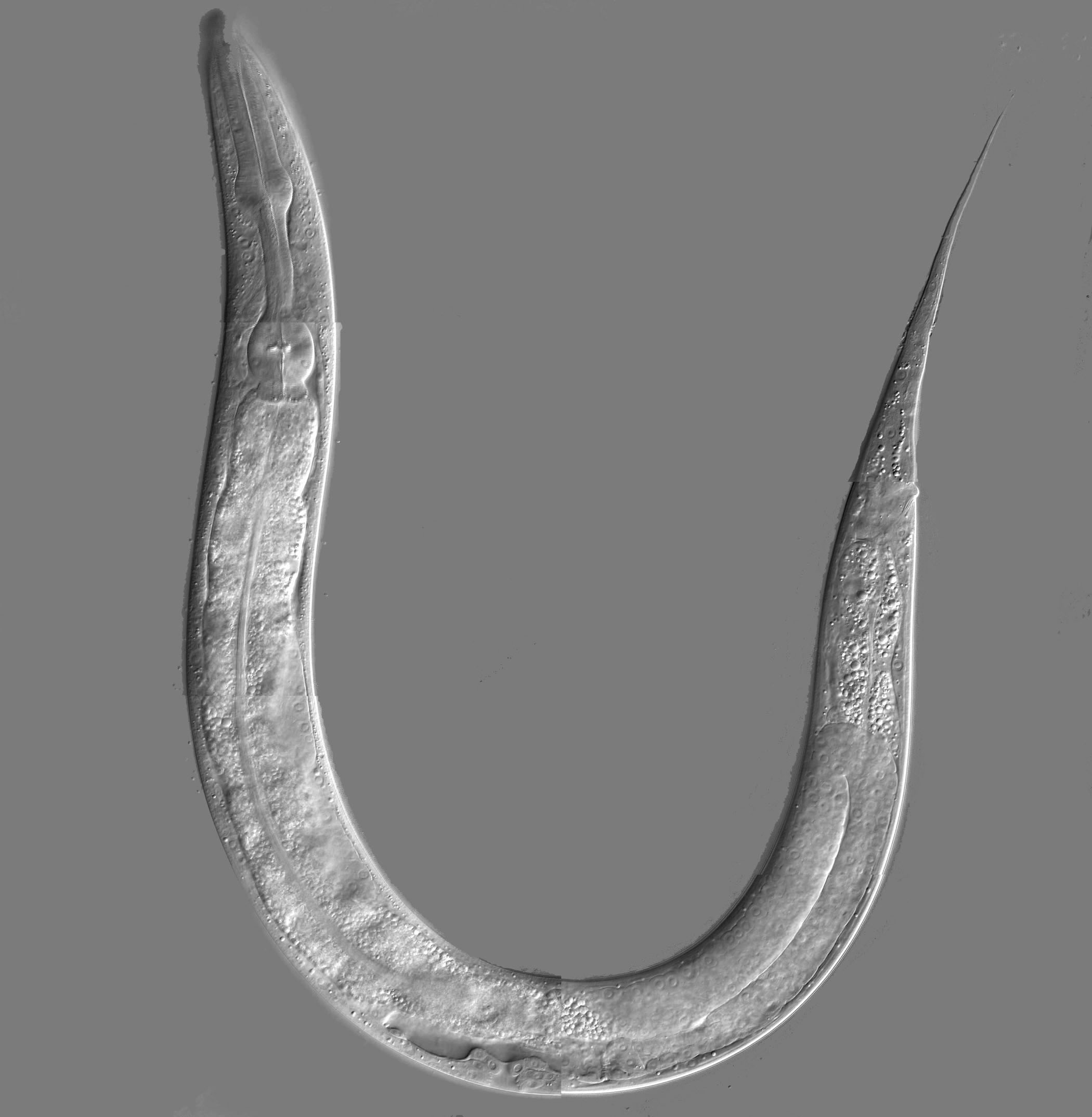
and her colleagues observed this marvel in a number of organisms such as plants, mice and even human diseases. In the plants they studied, it was linked to cell metabolism and in mice, it was linked to their brain function. This editing was also linked to ALS and epilepsy in humans. 

According to the article, Li and her colleagues also have been conducting a project known as the “1000 Genomes Project”, with a goal to reach the genomes of 1000 people. In this project, the researchers plan to compare the DNA and RNA sequences. “The results suggest that a vast amount of editing could be occurring across the genome, with the researchers identifying more than 102,000 potential editing events” (Translation). One thing the study suggested was that 97% of the gene transcripts are changed after a new template of RNA molecules has been formed from the DNA code. Li saw this rate as “surprisingly high” because normally in most cases of RNA editing, there were only 2 types of alteration, but many other differences that Li and her colleagues found are seemingly unknown. They also concluded that nobody can tell where the edited RNA is translated to. 

For Emmanouil Dermitzakis, he is more skeptical than Mingyao Li. He is also one of the researchers on the “1000 Genomes Project” and he argues DNA sequences of the mismatched pairs may simply be because of sequencing errors. He thinks that this editing varies between different cell types. The article states, “The only way to check would be to re-sequence a large number of the genes that seem to be edited.” This is being done by Li now, in hopes to be able to really prove this fascinating phenomenon. 

Blog post Author: Brooke Vasilescu Section 124-26

Work Cited:

Katsnelson, Alla. “DNA Sequence May Be Lost in Translation.” Nature.com. Nature
                       Publishing Group, 05 Nov. 2010. Web. 24 Feb. 2013.

“RNA Editing Definition-Medical Dictionary Definitions of Popular Medical Terms Easily
                       Defined on MedTerms.” Medterms. N.p., n.d. Web. 24 Feb. 2013. 

Wednesday, February 13, 2013

Will This Fish Transform Medicine

Photo courtesy of popsci.com
Researchers and geneticists looking for the cause of rare diseases and illnesses are now using the translucent zebra fish in replacement of rodents. The ability to mutate zebra fish DNA is allowing researchers to manipulate human diseases into the fish as a model. 

Families of infants with suspected genetic diseases or illnesses can send a vile of blood to the Human Genome Sequencing Center at the Baylor College of Medicine. There, researchers and scientists can accurately and quickly sequence the infant’s DNA and make copies. If a mutation is found in the infant’s DNA sequence, as suspected, researchers can steadily insert the same DNA mutations into the larval zebra fish. Then, through observance, researchers can watch for any structural defects that mature. Since the fish is transparent, organ development is easy to detect. For example, researchers watched a larva zebra fish grow a heart on the right side of its body, similar to the child with the DNA sequencing for this characteristic. Scientists were able to narrow down this child’s six mutations in her sequencing to the exact one relating to the heart displacement. 

Virginia Hughes, author of the original article, reports the zebra fish have been manipulated with “DNA mutations linked to narcolepsy, muscle disorders, and the large head size associated with autism.” Not only do researchers envision the zebra fish to display DNA mutations, but also as a test for treatment. The fish, unlike rodents, absorb drugs in the water through their skin. From this, scientists can observe which drugs and molecules affect the fish. Those drugs with hopeful effects are then tested in mammals such as rodents. Leonard Zon (of Harvard Medical School) discovered with one particular drug, the animals’ blood stem-cell counts amplified. This was then used for leukemia patients who had depleted blood cells from radiation treatment. The drug was about 83% effective for his twelve “test patients.” As one can see, zebra fish are advancing the medical field. Who would have thought such an unrelated animal to humans could be a source for medical mysteries and treatments.

Blog Post Author: Nicole Boisvert, Section 124-26
----------------------------------------------------------------------------------------------------
Work Cited
Hughes, Virginia. "Will This Fish Transform Medicine?" Popular Science. N.p., 21 Jan. 2013. Web. 11 Feb. 2013.


King Richard III Found!


Photi courtesy of montrealgazette.com

An astonishing discovery has recently occurred in a parking lot located in Leicester, England. Although this parking lot may not of been where Richard III - a past king of England who has been deceased for around 500 years prior to the discovery - would have wanted to be buried, it seems that this is where he was all these years. Genealogists proved that this body was indeed Richard III by comparing his DNA taken from the bones of skeletons found from deceased family members.
            Along with DNA testing, radiocarbon dating further proves that this is Richard III – as if DNA proof isn't enough! The test proves that the bones were present in the 15th to 16th century, which matches up with Richard’s timeline. Furthermore, the skeleton fits with the description that Richard III was said to have: lean, slender frame with a severe case of scoliosis.
            Curious of the poor man’s death? The bones show that Richard was most likely killed by two injuries that occurred to his skull, along with ten other wounds found along the skeleton, but the skull injury being the most likely cause of his death. It seems that his hands were tied and he was also humiliated by being stabbed in the buttocks at the time of his murder by some hooligans who jumped him in the middle of a street.
            All of the factors such a skeleton positioning, carbon dating, and most importantly DNA testing proves that this skeleton found buried under a parking lot in England has been harboring a past monarch of the great English nation for over 500 years. Fascinating discovery!


Blog Post Author: Kelsey Martin, Section 124-23

Citation
            McCarthy, Eugene. "King Richard III found!." . Macroevolution.net, 7 Feb 2013. Web. 11 Feb 2013. <http://www.macroevolution.net/richard-iii-found.html>

A Brave New World of Prenatal DNA Sequencing

Photo courtesy of bioedge.org
The leading DNA sequencing company, Illumina, is making moves to become a larger presence in the world of prenatal testing by recently acquiring the new company Verinata. The current procedures offered to mothers whose fetus's are at high risk for chromosomal abnormalities are very invasive and can cause miscarriages. Instead of withdrawing samples from the placenta and/or amniotic fluid, they wish “to search for these chromosome disorders starting from 10 weeks of gestation on, … [by] sequenc[ing] the bits of DNA floating in a sample of the mother’s blood” (Young, 6).

“Around 12% of the DNA in the mother's blood belongs to her fetus” (Young, 7). This is much less invasive and risky. Also it could be offered to all soon to be mothers instead of just those with high-risk pregnancies. They soon hope to be able to test for not just whole chromosomal errors, such as Down Syndrome or Edwards Syndrome which result from trisomy of chromosomes twenty-one and eighteen, respectively, but to be able to map the fetus's entire genome and find specific gene mutations and disorders. Right now the only thing blocking this from happening is money. “‘As sequencing gets cheaper and cheaper, if you wanted to look at a particular mutation, say, in the cystic fibrosis gene, you may be able to do that in the future,’” says Heath (Young, 16).

This of course brings up ethical issues. If parents are able to find out that their child will have a debilitating disorder some may chose to abort the pregnancy.  Many believe that this is unethical and that people should live out their lives the way their cards were dealt, even if the hand is not that good.  With these new scientific possibilities more ethical questions are being raised as well.

Blog Post Author: Dani Kaprielian, Section 124-25.

Article Citation:
Young, Susan. “A Brave New World of Prenatal DNA Sequencing.” MIT Technology Review. 30 January 2013. Web. February 2013. <http://www.technologyreview.com/news/510181/a-brave-new-world-of-prenatal-dna-sequencing/>

Sunday, January 27, 2013

Aquarius Survives

In 1986, the George F. Bond was built.  It is an underwater habitat that is owned by NOAA.  By 1992, after surviving Hurricane Hugo in the U.S. Virgin Islands, the habitat was renamed the Aquarius and was relocated to the Florida Keys where it currently resides. 
Credit:  Leonard Ho, AdvancedAquarist.com


The habitat is a research base for Aquanauts wishing to spend up to 10 days, 66 feet underwater.  Why is this important for research?  If one were to dive from the surface with a SCUBA tank to a depth of  66 feet, you'd have an hour before having to re-appear on surface.  Basing oneself in the Aquarius, at 66 feet, leaving the habitat in SCUBA allows for up to 9 hours of research before having to return to Aquarius.

This facility allows researchers a decent amount of dive time to explore the Floridian coral reef system.  Since coral reefs are an early indicator of environmental changes, studying their behavior is important.  Additional research has been conducted by NASA for their NEEMO program since 2001.

NOAA, along with the University of North Carolina at Willmington, were the main funding force behind Aquarius.  In September of 2012, with the recession taking effect in all sectors of society, Aquarius was cut from their budgets.  Approximately $3 million dollars were spent each year on maintaining it's structure from the effects of salt water, hurricanes and use as a research base.   For a short time it seemed the life of Aquarius had come to an end.

This month, Florida International University (FIU) has negotiated with NOAA and funding has resumed through a grant from NOAA with FIU taking over direction of the lab.  This is excellent news for the research community to continue use of the Aquarius for another year. 

Article Citation:

Lam, Brian. "The more-certain future of Aquarius, the last undersea science lab." BoingBoing. 23 January 2013.  Web. 27 January 2013. <http://boingboing.net/2013/01/23/the-more-certain-future-of-aqu.html>.

Morgan, Curtis.  "FIU to take over underwater lab in Keys." Miami Herald. 16 January 2013.  Web.  27 January 2013. <http://www.miamiherald.com/2013/01/15/3184148/fiu-to-take-over-underwater-lab.html>.