Zoologger: The Largest Arthropod to Prowl the Land: Samantha Hill

Among the beautiful beaches and gigantic waves in Palmyra atoll, just south of Hawaii, a big creature waits his chance to emerge as the sun goes down.

Jawless Vertebrae Had the Worlds Sharpest Teeth: Meghan Skura

The Conodont is recorded to have the sharpest teeth of any animal that ever lived, hoever its teeth are barely thicker than a human hair.

Ancient Creature was a Miniture Monster: Christina Vilkhovoy

A 500 million year old monster looking predator was discovered in the deposits of Canada.

Some Echinoderms Will Never Grow Up: Erica DeMello

In life, people think of being “immature” or not growing up as a bad thing. But in this case, for species like echinoderms, it can be advantageous.

500-Million-Year-Old Sea Creature Unearthed: Desiree Villeneuve

Scientists have discovered a 520-million-year-old preserved fossil of an arthropod known as a fuxhianhuiid.

Paradox Worm' Xenoturbella Bocki Lacks Brain and Sex Organs, But Could Be Mankind's 'Progenito:Dana Hebert

There is a high possibility that a paradox worm can be more related to humans that many other more advanced organisms.

Sunday, March 31, 2013

Reproduction and Lifespan are intertwined

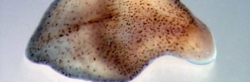
An organ-shaping gene has been found in the microscopic worm Caenorhabditis elegans, or C. elegans for short. The gene and the protein it makes are responsible for shaping the gonad, a complex reproductive organ. Similar genes and proteins are probably at work in other animals, including humans. The new finding by University of Wisconsin-Madison biochemist Judith Kimble and graduate student Robert Blelloch advances the prospect of one day growing complete organs for transplant.
Photo courtesy of Judith Kimble; news.wisc.edu

The gonad is well known for being helpful with reproduction but at the same time it has cut animals life spans down. Planck just recently discovered though that there is a molecular switch that can extend an animal’s life span using components of a developmental clock (Reproduction and Life Span Are Intertwined). The commonly used organism for research is called Caenorhabditis elegans. The Caenorhabditis elegans develops throughout four stages based on the developmental clock. “Yidong Shen and others worked with the Caenorhabditis elegans and used lasers to remove the germ cells, they found that remaining cells trigger the production of a hormone called Dafachronic acid (Reproduction and Life Span Are Intertwined).”

Dafachronic acid activates microRNAs, which work as little molecular switches causing changes in gene expression that promote longer life span. Losing the germ cells causes the developmental clocks to be put in motion leading to a longer life. I found this article intriguing because I never realized that research like this was going on, and that we have to ability to do such a research. It makes me want to continue looking up articles based on the theme of each lab to keep educating myself.

Blog Post Author: Paige Brandsdorfer Section 124-26

Works Cited
"Reproduction And Life Span Are Intertwined - ScienceNewsline." Reproduction And Life Span Are Intertwined - ScienceNewsline. N.p., n.d. Web. 10 Mar. 2013.
"Reproduction and Life Span Are Intertwined." ScienceDaily. ScienceDaily, 17 Dec. 2012. Web. 10 Mar. 2013.  

Clues to Nervous System Evolution Found in Nerve-Less Sponge

Sponges have all the genes necessary to have a nervous system, however they do not contain neurons at all. Research done on sponges has led to shocking discoveries of how the nervous system and gene expression are linked.
Photo courtesy of neurosciencenews.com.

It has always been question how sponges and animals have come from the same ancestry line but sponges lack the nervous system that animals have. Scientists realized that they could study the evolution of sponges to understand characteristics of the nervous systems in animals and the evolution of the nervous system.
            Sponges have all of the genes required to have the complex nervous system and create a synapse (mechanism that sends and receives signals), however, something happened over evolution that prevented sponges from having any neurons at all.
            Many scientists are interested in this idea and one scientist in particular, Danielle Bassett, decided to study sponge RNA and followed sponge activity. She looked at different stages of development in a sponge that lives on the Great Barrier Reef in Australia to analyze the genes that code for proteins that make up a synapse.
            What was found? Bassett found that the sponges would turn on and off unlike in animal cells that are either all on or all off.  This finding gave a good indication at the difference between animals and sponges, that sponges had cells that are not coordinated like animals. Sponges lacked the ability to express all the genes at once, as if they couldn’t be connected together. This prevented the creation of the synapse in the sponge. Therefore, throughout the evolution path, animals and sponges came from a common ancestry that had all the genes necessary to express a synapse, however animals split off because they have the ability to wire this network together.
            This discovery led to the idea of gene expression. Sponges lack the gene expression capabilities that animals have when it comes to neurons. Sponges can’t express these genes all at once but rather have an on an off thing going on. This research is just the beginning of much more information to be found about the evolution of the human brain!

Blog Post Author: Abby Mulligan Section 124-25 

Can a Jellyfish Unlock the Secret of Immortality?

In 1988 a marine biology student, Christian Sommer, discovered Turritopsis dohrnii, or more commonly known as the immortal jellyfish. Throughout his studies of this specimen he observed that the organism was incapable of death. The organism seemed to reverse its age until it reaches its earliest stage of life. 
Photo courtesy of the NewYorkTimes.com
In 1988 a marine biology student, Christian Sommer, discovered Turritopsis dohrnii, or more commonly known as the immortal jellyfish. Throughout his studies of this specimen he observed that the organism was incapable of death. The organism seemed to reverse its age until it reaches its earliest stage of life.

It is known that this rejuvenation is caused by environmental stress or physical assault. An unusual process, transdifferentiation, occurs within these organisms where one type of cell is converted into another. Turritopsis go through two main stages of life, the polyp and medusa. A polyp contains stalks that branch out and end in buds. Medusas are bell shaped with dangling tentacles. These characteristics describe most hydrozoans, yet the immortal jellyfish sink to the bottom of the ocean floor and the bodies fold in on themselves. The bell-dome reabsorbs the tentacles and continues to degenerate until it is ultimately a blob. An outer shell is formed over the gelatinous blob after a few days and root-like parts shoot out. These stolons lengthen and become polyps, which produce new medusas. An adult medusa is about the size of a fingernail.

The reverse aging process is not fully understood because it is difficult to attain good hydroid experts and the genus is difficult to culture in a laboratory. One consistent scientist, Shin Kubota, is dedicated to culturing polyps in his lab in Japan. Kubota said, “Once we determine how the jellyfish rejuvenates itself, we should achieve very great things. My opinion is that we will evolve and become immortal ourselves.”

It was only until recently that the ways of this jellyfish may be valuable in new discoveries with human immortality. Even though jellyfish have no brains, heart, and pass food and waste through the anus, it was found that both jellyfish and humans are very similar. Kevin J. Peterson, a molecular paleobiologist who worked with the study, said, “There’s a shocking amount of genetic similarity between jellyfish and human beings. From a genetic perspective, apart from the fact that we have two genome duplication  we look like a damn jellyfish.” More and more information about these miraculous creatures is being discovered, the relation to human beings cannot simply be certain until further research is accomplished.

Blog Post Author: Megan Giec from Section 124-21

Work Cited

Rich, Nathaniel. "Can a Jellyfish Unlock the Secret of Immortality?" The New York Times. N.p., 28 Nov. 2012. Web. 4 Mar. 2013. <http://www.nytimes.com/2012/12/02/magazine/can-a-jellyfish-unlock-the-secret-of-immortality.html?pagewanted=all&_r=0>.


Strep throat and OCD are linked, Israeli researchers find

Children suffering from strep throat should be closely monitored for symptoms of OCD, the researchers say.
Photo courtesy of Reuters

A group of researchers at Tel Aviv University recently discovered that an individual who has contact with the bacteria streptococcus A as a child can eventually lead to obsessive-compulsive disorder.  Although streptococcus A is commonly known as the bacteria that causes strep throat and minor pain, many people are not aware of the fact that it can ultimately develop into something more serious such as OCD. This article classifies OCD as “an anxiety disorder marked by repeated and intrusive thoughts that can trigger hording, obsessions and intensely repetitive behavior” (Even).

In this latest study, conducted by Prof. Daphna Joel (head of the psychobiological department in the Laboratory of Behavioral Neuroscience in the Department of Psychology at Tel Aviv University), rats displayed several characteristics and behaviors associated with OCD after being exposed to streptococcus A. The rats were first injected with streptococcus A microbes and then were given time to develop antibodies. After this, the rats then received an injection of a specific substance that enabled the antibodies to travel to their brains. Within the brain, the researchers found that the antibodies attached themselves to certain regions and also were “connected to changes in the level of neurotransmitters in the brain.”

In addition to the more complex changes in the brains of the rats, the infected rats also displayed altered “balance and coordination” and took part in obsessive grooming.  In addition, the researchers took this experiment one step further and were able to locate the specific dopamine receptors that the antibodies bound to. According to this article, such a discovery “will likely aid in development of treatments for OCD.” Scientists have been studying this “link” for over 20 years and Joel declares that “the description of the model in rats is of great significance.” This new evidence found in the rats is certainly a step in the right direction.   

Blog Post Author: Vickey Cooley Section 124-26 

Works Cited:
Even, Dan. "Strep Throat and OCD Are Linked, Israeli Researchers Find." Haaretz.com. Haaretz Daily Newspaper Ltd., 08 Jan. 2013. Web. 01 Mar. 2013.

Puddles of Goo? Brainless Slime Molds Have Memories


The yellow slime mold leaves a thick mat of translucent slime (left on the agar plate) behind it as it moves, ooze that it later avoids. Now researchers have found the goo uses the gel trail as a kind of memory to navigate. 
CREDIT: Image courtesy of Audrey Dussutour                                           


Scientists have found that just because an organism does not have a brain, does not mean that it cannot remember things. Slime molds, which were once considered fungus and later put into the protists classification, have been investigated in order  to see if they truly do have memories. Although it may not be the same definition of intelligence we think of inside of the human brain such as having the ability to create, store, and recall memories, this slime mold uses a different technique in order to remember where it has been. Slime molds do not have brains or even neurons; however, who needs either of those when you have ooze to lie down and retrace your steps.

This type of slime mold, also known as Physarum polycephalum, “leaves a thick mat of translucent slime behind it as it moves” (Choi). The slime mold can detect it’s own trail and use this as form of memory. It is almost like allowing itself “to communicate with it’s future self” (Choi). When the scientists tested this theory, slime mold that was able to detect it’s own trail reached the food placed in a maze 30% faster while slime that was blinded to their own trail took up to 10 times longer looking in places it had already visited. This was the first tested evidence that supported “a spatial memory system in a creature without a brain” (Choi).

Blog Post Author: Tyler Davis Section 124-26

Work Cited
Choi, Charles. "Puddles of Goo? Brainless Slime Molds Have Memories." LiveScience. N.p., 8 Oct. 2012. Web. 3 Mar. 2013. <http://www.livescience.com/23797- brainless-slime-mold-memories.html>.

Sunday, March 3, 2013

Discovery Of Giant Roaming Deep Sea Protist Provides New Perspective On Animal Evolution

Tracks of the Bahamian Gromia sphaerica(A) Gently sloping seafloor with numerous G. sphaerica visible. White arrowheads indicate notably curved tracks; black arrowheads indicate adjacent tracks running in opposite directions.(B) Alignment of the tracks on a steeper slope.(C–F) Details of the tracks, demonstrating the characteristic bilobed profile with the central ridge that is especially prominent near the organism. In panel (E), note that the track proceeds through a dip in the terrain, suggesting active locomotion. In panel (F), a group of three large cup corals growing on a half-buried sea urchin test indicate a remarkable sediment stability that may facilitate track persistence. Photos courtesy of sciencedirect.com.

New evidence about protists has led to the disproval of our current understanding of the evolution of the kingdom Animalia. A Biologist, Mikhail Matz, from the University of Texas at Austin was researching the ocean floor of the Bahamas when he came upon an amazing discovery. He found grooves on the deep floors of the ocean that came from protists, but showed a bi lateral pattern that is considered to be a trait of animals.

This is so incredible because it is the first time bi-lateral animal-like traces are appearing in single celled organism’s movements. The connection comes from the fact that the Precambrian era multi-cellular animals have very comparable trails left behind due to movement. “Bilateria appeared in the fossil record in the early Cambrian about 542 million years ago” (Science Daily). Mats says, “if our giant protists were alive 600 million years ago and the track was fossilized, a paleontologist unearthing it today would attribute it to a kind of large, multi-cellular, bilaterally symmetrical animal…We now have to rethink the fossil record” (Mats).  It is an amazing thing to be able to add knowledge to the pool of evolutionary theory.

External Appearance of the Bahamian G. sphaerica(A) Collected specimen demonstrating transparent membranous test, multiple evenly scattered apertures, and dark-green sediment contained in the protoplasm under the test's surface.(B) Freshly collected specimen with collapsed pseudopodia still visible (white projections).(C) Typical grape-shaped specimen in situ, fully covered with sediment. The track of this one is toward the top right corner of the picture.
Since fossils are a customary means of examining organisms, it is hard to be one hundred percent positive about this evidence because it is hard to extract a fossil from the ocean floor due to water movements. Fortunately for this protest, it’s location, with little current, made it possible to maintain the tracks. With such evidence it is now evident that the fossil record must be re examined to fit these protists in their appropriate location.

Blog Post Author: Jonathan Zirna Section 124-26


Works Cited
University of Texas at Austin (2008, November 21). Discovery Of Giant Roaming Deep Sea                     Protist Provides New Perspective On Animal Evolution. ScienceDaily. Retrieved March   3, 2013, from http:wwwsciencedaily.com/releases/2008/11/081120130531.htm

New Insights Into Protein Synthesis and Hepatitis C Infections

From left, Jennifer Doudna, Bunpote
 Siridechadilok and Eva Nogales, used this cryo 
electron microscope to create a 3-D model of
 the protein complex eIF3 that shed new light 
on protein synthesis and Hepatitis C viral

 infections. Photo courtesy of lbl.gov
This article shows how cryo electron microscopy (cryo-EM) was used to create a 3-D model of the protein complex called eukaryotic translation initiation factor 3 (eIF3).  The cryo-EM reconstructions of Biochemist Jennifer Doudna and biophysicist Eva Nogales show that eIF3 consists of five lobes; a head and a pair of arms and legs. 

The study shows that the left “arm" of the eIF3 complex binds to the eukaryotic protein complex that recognizes the methylated guanosine cap at the 5’end of the eukaryotic mRNAs.  By drawing the mRNA’s 5’end cap through the ribosome entry site and towards the exit, eIF3 ensures the mRNA is properly positioned for its genetic code to be translated. EIF3 will also wrap its arms and legs around a structural element of RNA for the hepatitis C virus (HVC), known as the internal ribosome entry site, and pin it to the exit site of the 40S ribosome subunit. The IRES leaves through the left arm of the eIF3 complex at the same location where interaction with the human mRNA cap-binding complex takes place. 

This act might explain the ability of the HVC IRES to hijack the human ribosome and its associated translation factor. This new information gives insight into how the initiation mechanisms of protein synthesis work specifically for humans. It also provides a step towards understanding what happens when a viral infection occurs at the molecular level. By understanding these methods, scientists may be able to come up with new and improved therapies for viral infections.

Blog post Author: Jenn Gregorio Section 124-26

Work Cited:
"New Insights Into Protein Synthesis and Hepatitis C Infections." Research News:. N.p., 5 Dec. 2005. Web. 03 Mar. 2013. 

DNA sequence may be lost in translation

Photo courtesy of  wired.com


A new mysterious, unexpected phenomenon was brought to the attention to the Annual Meeting of the American Society of Human Genetics in Washington, DC. This phenomenon is called “RNA editing.”  RNA editing is, “The process by which messenger RNA is modified (edited) after it is synthesized before it is translated into protein” (MedTerms). We all know that DNA is transcribed first to form RNA and then what codes for the amino acids depends on the triplets of RNA. The RNA sometimes “edits” for different bases, switching them, hence the name “RNA editing.” Two studies, conducted by Mingyao Li and Emmanouil Dermitzakis presented at the meeting suggested and showed many different opinions and findings. 

Li and her colleagues observed this marvel in a number of organisms such as plants, mice and even human diseases. In the plants they studied, it was linked to cell metabolism and in mice, it was linked to their brain function. This editing was also linked to ALS and epilepsy in humans. 

According to the article, Li and her colleagues also have been conducting a project known as the “1000 Genomes Project”, with a goal to reach the genomes of 1000 people. In this project, the researchers plan to compare the DNA and RNA sequences. “The results suggest that a vast amount of editing could be occurring across the genome, with the researchers identifying more than 102,000 potential editing events” (Translation). One thing the study suggested was that 97% of the gene transcripts are changed after a new template of RNA molecules has been formed from the DNA code. Li saw this rate as “surprisingly high” because normally in most cases of RNA editing, there were only 2 types of alteration, but many other differences that Li and her colleagues found are seemingly unknown. They also concluded that nobody can tell where the edited RNA is translated to. 

For Emmanouil Dermitzakis, he is more skeptical than Mingyao Li. He is also one of the researchers on the “1000 Genomes Project” and he argues DNA sequences of the mismatched pairs may simply be because of sequencing errors. He thinks that this editing varies between different cell types. The article states, “The only way to check would be to re-sequence a large number of the genes that seem to be edited.” This is being done by Li now, in hopes to be able to really prove this fascinating phenomenon. 

Blog post Author: Brooke Vasilescu Section 124-26

Work Cited:

Katsnelson, Alla. “DNA Sequence May Be Lost in Translation.” Nature.com. Nature
                       Publishing Group, 05 Nov. 2010. Web. 24 Feb. 2013.

“RNA Editing Definition-Medical Dictionary Definitions of Popular Medical Terms Easily
                       Defined on MedTerms.” Medterms. N.p., n.d. Web. 24 Feb. 2013.